|
我们的研究与国外的研究结果相似,均表明OCT能较准确地在活体上测量RNFL厚度,由于它是一种非接触性、非损伤性、利用光进行高分辨率的影像学检查,所用的是近红外光,患者几乎觉察不到,而其背景光为红色光,因而患者可以很好地配合和耐受检查,即使是10岁左右的儿童也可以很好地配合检查,因而可获得较满意的图像进行分析。但由于OCT是利用光进行检查,因而明显的屈光间质混浊将对检查结果,尤其是精确的定量检测结果产生一定的影响。但无论如何OCT仍是一种精确测量RNFL厚度的方便、快捷方法,在对评价各种疾病的RNFL方面有较重要的临床价值。

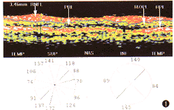
图1 正常人视乳头周围视网膜OCT图像,RNFL示视网膜神经纤维层(红色反光) pRL示光感受器细胞层(暗区) ,I 和OPL示内外丛状层(黄绿相间),RPE示视网膜色素上皮 (红白色反光) 。 TEMP示颞侧,SUP示上方,NAS示鼻侧,INF示下方。左侧圆形方位图示各钟点RNFL厚度均值,右侧圆形方位图示 4个象限平均RNFL厚度值 (左为颞侧)

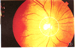
图2 正常人眼底, 圆圈为扫描方式, 直径3.46mm,箭头示扫描方向
基金项目:广东省科委重点基金资助项目(49)
作者单位:刘杏(510060广州,中山医科大学中山眼科中心)
凌运兰(510060 广州,中山医科大学中山眼科中心)
骆荣江(510060 广州,中山医科大学中山眼科中心)
葛坚(510060 广州,中山医科大学中山眼科中心)
周文炳(510060 广州,中山医科大学中山眼科中心)
郑小平(510060 广州,中山医科大学中山眼科中心)
参考文献
1,Hoyt WF, frisen L , Newman NM. Funduscopy of nerve fiber layer defects in glaucoma. invest Ophthalmol Vis Sci, 1973, 12: 814-829.
2,Airaksinen PJ , Nieminen H . Retinal nerve fiber layer photography in glaucoma. Am J Ophthalmol, 1985, 92: 877-879.
3,Weinreb RN , Shakiba S , Zangwill L . Scanning laser polarimetry to measure the nerve fiber layer of normal and glaucomatous eyes. Am J Ophthalmol,1995, 119: 627-636.
4,Anton A , Zangwill L , Emdadi A, et al . Nerve fiber layer measurements with sanning laser polarimetry in ocular hypertension. Arch Ophthalmol, 1997,115: 331-334.
5,Schuman JS , Hee MR , Puliafito CA, et al . Quantification of nerve fiber layer thickness in normal and glaucomatous eyes using optical coherence tomography. Arch Ophthalmol, 1995, 113: 586-596.
6,Quigley HA, Addicks EM . Quantitative studies of retinal nerve fiber layer defects. Arch Ophthalmol, 1982, 100: 807-814.
7,迟启民,富田刚司,北克明,等.应用激光偏光扫描测量仪评价正常人视网膜神经纤维层厚度与年龄的关系.中华眼科杂志,1998,34:199-201.
8,Schuman JS, Pedut-Kloizman T, Hertzmark E, et al. Reproducibility of nerve fiber layer thickness measurements using optical coherence tomography. ophthalmology, 1996, 103:1889-1898.
上一页 [1] [2] [3] |
