中华眼科杂志 1999年第6期第35卷 眼底病
作者:魏文斌 杨文利 赵丽丽 史翔宇 陈铮 王景昭
单位:100730 首都医科大学附属北京同仁医院眼科
关键词:光学相干断层成像;黄斑裂孔
【摘要】 目的 探讨黄斑裂孔的光学相干断层成像(optical coherence tomography, OCT) 特征及OCT临床应用价值。方法 1998年9~12月临床诊断为黄斑裂孔者共35例(38只眼)。经双眼散瞳后进行OCT检查,对获取的图像进行分析和测量。结果 1例(1只眼)OCT显示为玻璃体黄斑牵引;1例(1只眼)为黄斑前膜所致的假性裂孔;33例(36只眼)为黄斑裂孔,其中3例累及双眼。36只眼中,板层黄斑裂孔4只眼,OCT图像表现为黄斑中心窝处神经上皮部分缺失,但无神经上皮脱离晕轮,未见神经上皮层间的小囊泡;全层黄斑裂孔32只眼,OCT图像特征为黄斑中心窝处可见边界清晰、锐利的视网膜神经上皮全层缺失,裂孔周围有神经上皮脱离的晕轮,部分患者的神经上皮间无反射的小囊腔,裂孔周边视网膜增厚。特发性黄斑裂孔26只眼, 按Gass分期,属于I期裂孔2只眼,II期裂孔3只眼,III期裂孔15只眼,IV期裂孔6只眼。4只眼经玻璃体手术治疗后OCT图像显示裂孔闭合,裂孔周边晕轮消失。 定量测定裂孔直径为(565.88±40.35)μm,裂孔周围晕轮直径为(1338.76±147.57)μm。裂孔边缘视网膜厚度为(391.87±18.97)μm。经Pearson相关性分析,裂孔大小、晕轮范围、裂孔边缘视网膜厚度均与视力相关。结论 OCT是一种新的非接触性,非侵入性光学断层检测技术,对黄斑裂孔的诊断、鉴别诊断、定量评估、病情监测、术式选择、疗效评价等方面具有重要的临床应用价值。
Optical coherence tomography of macular holes
WEI Wenbin, YANG Wenli, ZHAO Lili, et al. Department of Ophthalmology, Beijing Tong Ren Hospital, Beijing 100730
【Abstract】 Objective To study the characteristics and clinical application value of the optical coherence tomography (OCT) of macular holes. Method A total of 35 patients with the clinical diagnosis of macular hole were examined with OCT between September and December 1998. OCT imaging was conducted through a dilated pupil, and the OCT images were analyzed and measured. Results Of the 35 patients examined with OCT, there were pseudohole and epimacular membrane in one eye, vitreofoveal traction in one eye, macular holes in 36 eyes, and 3 patients had macular hole in bilateral eyes. In 4 eyes, there were partial-thickness macular holes, and the defect of partial thickness of neural epithelium in the fovea without halo of retinal detachment was shown in the OCT image. In 32 eyes, there were full-thickness holes; the OCT displayed complete losing of the whole thickness of the neural epithelium in the fovea, sharp edge of the hole and the halo of retinal detachment around the hole. Sometimes nonreflective cavities could be seen within the retina, and the retinal thickness around the hole was increased. According to Gass stage classification of macular hole, there were 2 eyes with impending hole, 3 eyes in stage 2, 15 eyes in stage 3 and 6 eyes in stage 4. 4 eyes underwent vitrectomy. The OCT imaging after the surgery demonstrated the closure of hole and the disappearance of halo surrounding the hole. Through quantitative measurement, the diameter of the hole was (565.88±40.35)μm, the diameter of the halo was (1 338.76±147.57) μm, and the retinal thickness surrounding the hole was (391.87±18.97) μm. The sizes of the hole and the halo and the retinal thickness around the hole were correlated with the vision. Conclusion OCT is a novel noninvasive, noncontact imaging technique. It is helpful in the diagnosis and differential diagnosis of the macular hole; the progress of the hole can be quantitatively estimated, and it is also helpful in selection of operation and assessment of operative therapeutic effects.
【Key words】 Optical coherence tomography Macular hole
光学相干断层成像(optical coherence tomography, OCT) 是一种新的非接触性,非侵入性光学检测技术,可对眼组织进行断层成像,垂直分辨率高达10μm,对黄斑疾病的诊断、鉴别诊断、病情监测及定量评估等方面具有临床实用价值[1-3]。黄斑裂孔是常见的眼底疾患,应用眼底生物显微镜、眼底血管荧光造影及眼底照相等检查方法难以避免误诊、漏诊,且对其发病机制研究、治疗方法选择等方面的作用有限。本文旨在研究黄斑裂孔的光学相干断层成像特征及其临床应用价值。
资料与方法
1.病例选择:1998年9~12月在我院就诊的35例(38只眼)黄斑裂孔患者,经询问病史,一般眼科常规检查,直、间接检眼镜及裂隙灯显微镜检查眼底,部分经眼底血管荧光造影、眼底摄影、视野检查,经临床确诊为黄斑裂孔者。其中男性12例,女性23例;年龄20~81岁,平均56.8岁。患者均因视物变形及视力减退就诊。6例有眼球钝挫伤史,余29例无明显诱因,无其他眼疾史。38只眼的视力均为0.01~0.6,平均0.095±0.023;其中7只眼为-1.0~-6.0 D近视,31只眼的屈光度在±1.0D以内。
2.检查方法:受检者双眼充分散瞳,保持坐位,调整仪器,选择线性扫描,获得满意图像后贮存,对图像进行分析及定量测定。
3.统计学分析:将所测数据用SPSS软件包进行Pearson相关性分析。
结果
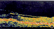
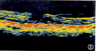
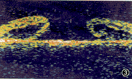
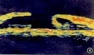
1.黄斑裂孔的OCT图像特征:35例(38只眼)临床诊断为黄斑裂孔的患者, 经OCT检查,1例(1只眼)显示为玻璃体黄斑牵引(图1),1例(1只眼)为黄斑前膜所致的假性黄斑裂孔(图2)。33例(36只眼)黄斑裂孔中,有3例双眼受累,1例对侧眼可见玻璃体后脱离。 全层黄斑裂孔32只眼的OCT图像特征:黄斑中心窝处可见边界锐利、清晰的视网膜神经上皮层的全层缺失,裂孔周围有神经上皮脱离的晕轮,14只眼可见神经上皮层间无反射的小囊泡(图3),4只眼可见裂孔的盖膜(图4),6只眼可见脱离的玻璃体后界膜(图5),6只眼可见玻璃体黄斑牵引(图6)。板层黄斑裂孔4只眼的OCT图像特征:中心窝处神经上皮层部分缺失,无神经上皮脱离的晕轮,亦未见神经上皮层间的小囊泡(图7),1只眼可见黄斑部玻璃体牵引。

图1 患者女, 60岁, 右眼视物变形3个月, 视力0.3。OCT示黄斑部视网膜增厚,可见微囊样改变及玻璃体黄斑牵引

图2 患者女, 58岁, 右眼视力减退伴视物变形1年, 视力0.6。OCT示黄斑部玻璃体视网膜牵引, 前膜形成, 视网膜感觉层未缺失

图3 患者男, 72岁, 右眼视力减退半年, 视力0.1。OCT示黄斑神经上皮全层缺失, 裂孔周围视网膜增厚, 并可见微囊样改变

图4 患者女, 67岁, 左眼视力减退, 视物变形3个月, 视力0.2。 OCT示黄斑区神经上皮全层缺失, 裂孔周围视网膜增厚, 裂孔前玻璃体可见裂孔盖膜
[1] [2] 下一页 |
