眼科研究 2000年第1期第18卷 临床研究
作者:王福 张奎启
单位:大连医科大学口腔解剖基础教研室 116027
关键词:颈内动脉;眼动脉;脑膜中动脉
摘要 目的研究眼动脉的起始、走行、分 支和分布,为眼科临床提供参考。方法对23例成人头颅标本眼动脉进行解剖。结果 66.7%的眼动脉起自颈内动脉,33.3%的眼动脉来自颈内动脉和脑膜中动脉并相互吻 合。其主要分支包括睫状后动脉、视网膜中央动脉、泪腺动脉等。结论眼动脉主要来自 颈内动脉和脑膜中动脉,视网膜中央动脉全部来自颈内动脉,表明视网膜的血供与颅内血供密切相关。
分类号 R361.1
An atomical observation of ophthalmic arter y and branches
Wang Fu Zhang Kuiqi
(Depa rtment of Stomatology,Dalian Medical Uni versity,Dalian116027)
Abstract Objectiv eTo provide anatomic data basis for cli n ic ophthalmology,the origin,course,branc hes and distributions of ophthalmic arte ry were further investigated.Methods23 skulls of human cadavers were irrigated with red latex and fixed in formalin.The n the roof of the orbit were opened and 45 ophthalmic arteries were carefully st u died within the orbit.Some arteries were measured.Results67.7% ophthalmic arte ry arose from the internal carotid artery, 33.3% ophthalmic artery arose from both the internal carotid artery and the midd le meningeal artery,but in two cases the main distribution of blood came from th e middle meningeal artery.According to t he relation to the optic nerve,the intra -orbital course of ophthalmic artery wer e divided into three parts.The main branc hes of ophthalmic artery included poster i or ciliary artery,central retinal artery ,lacrimal artery.ConclusionThe ophthal mic artery mainly comes from the interna l c arotid artery and the middle meningeal a rtery.Most of the origin site of branche s were located on the “Angle”,the second part and “Bend” in the ophthalmic arter y.All the central retinal arteries comin g from internal carotid artery indicated bloo d supply of retina is closely related to blood supply of intra-cranium.
Key words internal carotid artery ophthalmic art ery mid dle meningeal artery
关于眼动脉的解剖Hayreh等曾做了详尽地报道[1~5],国内报道不多[6,7],且各家观点不完全一致,本文解剖了45支眼动脉。
1 材料与方法
用红色乳胶灌注的福尔马林固定的成年头颅23例(22例双侧解剖,1例只解剖1侧),去脑凿去眶上壁显示眼动脉,观察其起始、走行及其分支。用游标卡尺测量有关距离和动脉管径。
2 结果与讨论
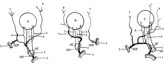
2.1 眼动脉的起始:本文45支眼动脉,其中左侧22支,右侧23支。均起自颈内动脉前床突部。其中15支与脑膜中动脉有交通(图1A),有2支以脑膜中动脉为主(图1C)。

图1 眼动脉及泪腺动脉的来源
Fig.1 Variati ons in the origin of ophthalmic artery a nd lacrimal aretry
1.颈 内动脉 2.脑膜中动脉 3.眼动脉 4.颞侧睫状后动脉 5.鼻侧睫状后动脉 6.视网膜中央动脉 7.泪腺动脉 8.眶上动脉OC:视神经 管 ON:视神经 SOF:眶上裂 1.Internal carotid arte ry 2.Ophthalmic artery 3.Middle meningea l artery 4.Lateral posterior ciliary art ery 5.Medial posterior ciliary artery 6. Central retinal artery 7.Lacrimal artery 8.Supra-orbital artery OC:optic canal O N:optic nerve SOF:superior orbital fissu re
眼动脉与视神经共同包于视神经的硬膜鞘内,通过视神经管后入眶。在视神经管内有33支眼动脉在视神经的下外方(73.3%);有12支眼动脉在视神经的下方(22.7%)。未见有眼动脉经眶上裂入眶的(Hayreh[1]所述经眶上裂入眶)。用游标卡尺测量了眼动脉的管径,平均(1.40±0.27)mm。
2.2 眼动脉的眶内行程:根据眼动脉与视神经的关系,眼动脉可分为3段:第1段位于视神经下外侧;第2段在视神经上方或下方横过视神经;第3段在视神经内上方前行。眼动脉在行程中2次转变方向形成2个弯曲,1,2段之间的弯曲称角(angle);2,3段之间的弯曲称弯(bend)[2]。
2.2.1 眼动脉的第1段:眼动脉出视神经孔后至横过视神经之前,眼动脉位于视神经下外方的42支(93.3%);位于下内方的2支(4.4%);位于下方的1支(2.2%)。
2.2.2 眼动脉的角:多位于视神经外侧,锐角9支,占20%;钝角16支,占35.6%;直角20支,占44.4%。眼动脉的角多为钝角和直角。该结果与Hayreh[1]一致,与崔模[6]结果相反。
2.2.3 眼动脉第2段:此段横过视神经斜向前,其中行于视神经上方的39支,占86.7%;行于视神经下方的6支,占13.3%。这与Hayreh[2]、崔模[6]的结果相似。张诗兴[7]报道在49支中仅有1支行于视神经下方。
2.2.4 眼动脉的弯:位于2,3段之间,与视神经内上侧,不与视神经接触,多呈钝角。
2.2.5 眼动脉第3段:位于视神经内侧,在内直肌和上斜肌之间下,延续为滑车上动脉和鼻背动脉,仅有1例终止在筛前动脉。此段走行极度弯曲以适应眼球的运动[4]。
2.3 眼动脉的主要分支:眼动脉按所供应的区域划分为:眶组(泪腺动脉、眼肌动脉);眼球组(视网膜中央动脉、睫状动脉);眶外组(筛前后动脉、眶上动脉、睑内侧动脉、鼻背动脉、滑车上动脉)。按起始顺序分为:视网膜中央动脉和/或颞侧睫状后动脉、眼肌支、泪腺动脉和鼻侧睫状后动脉、筛后动脉、筛前动脉。
2.3.1 视网膜中央动脉
视网膜中央动脉起于眼动脉第1段者40支(88.9%);起于第2段者5支(11.1%)。起于第1段者多位于角处,其中单独起于角处25支,与鼻侧睫状后动脉共干的11支,与颞侧睫状后动脉共干的3支,与下肌支共干的1支;起于第2段者与鼻侧睫状后动脉共干的2支,与下肌支共干的1支。这一结果与崔模[6]相似。
视网膜中央动脉发出后迂曲前行,在视神经下面中线偏内侧穿入视神经的占77.8%;偏外侧穿入视神经的22.2%。这与崔模[6]结果相似,张诗兴[7]报道视网膜中央动脉多在视神经下方穿入,且发现有2支视网膜中央动脉穿入视神经。入视神经处与眼球后极的距离见表1,其管径值见表2。
表1 视网膜中央动 脉穿入视神经处 至眼球后极的距离(mm)
Tab.1 Dis tance between the point which the retina lcentral artery
pierces the optic nerve and the ocular posterior pole(mm)
| |
n |
Max |
Min |
 ±s ±s |
| OS |
22 |
11 |
5.4 |
8.36±1.63 |
| OD |
23 |
11 |
4.0 |
8.23±2.25 |
| total |
45 |
11 |
4.0 |
8.27±1.93 |
表2 视网 膜中央动脉外径(mm)
Tab.2 The outside diameter of retinal central artery(mm)
| |
n |
Max |
Min |
 ±s ±s |
| OS |
22 |
0.8 |
0.3 |
0.58±0.14 |
| OD |
23 |
0.6 |
0.3 |
0.47±0.09 |
| Total |
45 |
0.8 |
0.3 |
0 .57±0.13 |
无论眼动脉起自颈内动脉或脑膜中动脉,视网膜中央动脉100%来自颈内动脉,这说明视网膜中央动脉的血供与颅内血供密切相关。
2.3.2 睫状后动脉:多以2~3支起自眼动脉角处。本文共有睫状后动脉101支;鼻侧睫状后动脉43支,颞侧睫状后动脉47支,上睫状后动脉1支,起于下肌支的10支。其中有2条的34例(75.6%)(图1A,B,C);有3条的11例(24.4%)。有2条者皆为鼻侧和颞侧各1条睫状后动脉;有3条者每例为鼻侧1条睫状后动脉,颞侧2条睫状后动脉。另外有少数睫状后动脉(10支)起自肌动脉,其中有1支为上睫状后动脉[1,6,7]。
睫状后动脉发出后平行于视神经前行,在眼球后发出数支,其中1支睫状后长动脉和数支睫状后短动脉进入眼球。睫状后动脉走行分布大致可分为3种类型:Ⅰ型:睫状后动脉沿视神经先向下然后垂直向上分成数支进入眼球;Ⅱ型:睫状后动脉沿视神经先向上然后垂直向下分成数支进入眼球;Ⅲ型:睫状后动脉沿视神经水平直向前分成数支进入眼球[5]。
2.3.3 泪腺动脉:来源大致有3种,一种是完全起自脑膜中动脉(16支,35.6%)(图1B);第二种是完全起自眼动脉(19支,42.2%);另一种是起自前2者,以脑膜中动脉为主(10支,22.2%)(图1A,C);泪腺动脉发出后,向前外方走行于眼外直肌上缘,多以2支分布于泪腺。
2.4 眼动脉的其它分支:包括眶上动脉、滑车上动脉、鼻背动脉、筛前动脉、筛后动脉,以及至眼外肌的小动脉。
1,Hayreh SS, Dass R.The ophthalmic artery:Ⅰ.Origin an d intra-cranial and intra-canalicular co urse.Br J Ophthalmol,1962,46∶65
2,Hayreh SS,D ass R.The ophthalmic artery:Ⅱ.Intra-orbi tal course.Br J Ophthalmol,1962,46∶165
3,Hayreh SS.The ophthalmic artery:Ⅲ.Branc hes.Br J Ophthalmol,1962,46∶212
4,Hayreh SS.Artery of the orbit in the human bei ng.Br J Surg,1963,938
5,Yoshii I,Iked a A.A new look at the blood supply of th e retroocular space.The Anatomical Recor d,1992,233∶321
6,崔模,魏宝林,柴戢臣,等.眼动脉及其主 要分支的 研究.中华眼科杂志,1984,20∶30
7,张诗兴,侯守仁.颈内动脉前床突上段 与眼动脉的显微外科解剖.解剖学报,1986,127∶131
收稿:1999-02-06
修回:1999-09-08 |
