|
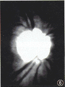
图6 采用阈值分割法后,视盘颞下方的RNFL反光消失,上,下方的RNFL分布不对称
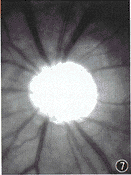
图7 青光眼的RNFL图像不清晰
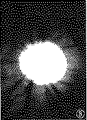
图8 采用阈值分割法后,视盘上方的RNFL反光首先消
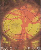
图9 高度近高眼视盘周围萎缩环
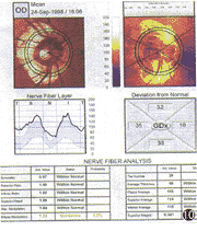
图10 NFA检测结果显示其伪信号明显增强
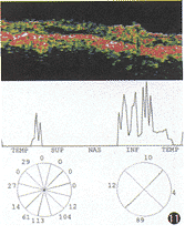
图11 OCT检测结果显示RNFL信号明显衰减
参考文献
1,Sommer A, Miller Nr, Pollack I, et al. The never fiber layer in the diagnosis of glaucoma. Arch Ophthalmol, 1977, 95: 2149-2156.
2,Sommer A, Katz J, Quigley HA, et al. Clinically detectable nerve fiber atrophy precedes the onset of glaucomatous field loss. Arch Ophthalmol, 1991, 109: 77-83.
3,Quigley HA, Dunkelberger GR, Green WR. Retinal ganglion cell atrophy correlated with automated perimetry in human eyes with glaucoma. Am J Ophthalmol, 1989,107: 453-464.
4,Greenfield DS. Optic nerve and retinal nerve fiber layer imaging in glaucomatous optic neuropathy. Int Ophthalmic Clin, 1999, 39: 121-145.
5,Tuulonen A, Alanko H, Hyytinen P, et al. Digital imaging and microtexture analysis of the nerve fiber layer. Glaucoma , 2000, 9: 5-9.
6,Weinreb RN, Shakiba S, Zangwill L. Scanning laser polarimetry to measure the nerve fiber layer of normal and glaucomatous eyes. Am J Ophthalmol, 1995,119: 627-636.
(收稿日期:2000-04-26) 上一页 [1] [2] [3] |
