|
�������ڻ��߹�26����9������ɸ������5������ɸ�����8������ɸ��ǰ����4����������6���⣬����20�������������ơ�����3��������2������ɸ������(����������)��1����������(δ��������)��
����291����������5������������ϸ���ֻ�������������3��PEC-stage�����(2���������������϶˵�����ϸ������)��1��Ϊ˫�ۻ��ߣ�һ��ժ������һ���з������ƣ�ժ���۵�PEC-stageΪ0�ڣ�����ɸ��ǰ�������������۵����������������ѹ���ߣ��������̷����Լ�Һ��������ϸ������4����������ϸ����ת����������5��Ϊ˫��RB���ߣ�PEC-stageΪ���ڣ�����Ҳ��ɸ��ǰ����������ϵ�����˵ڶ����������ܰ�ϸ����Ѫ�������ں͢�����һ����������������������RB��ϸ��ת�Ƶ��µ���������0������������1������ת����Ϊ0.4%������������3������ת���ʴ�12.0%��
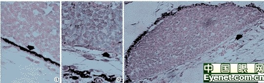
����ͼ1������Ĥɫ����Ƥ��Ӧǰ��(0��)������Ĥɫ����Ƥ����(��)��HE��400��ͼ2������Ĥɫ����Ƥ��Ӧ��(��a��)������ϸ��(RB)�벣��Ĥֱ�ӽӴ����ô�������Ĥɫ����Ƥ��ʧ������Ĥ����(��)��PAS��400��ͼ3������Ĥɫ����Ƥ��Ӧ��(��b��)������Ĥɫ����Ƥ(RPE)��ʾ��Ӧ�Ա仯���벣��Ĥ���벢¡������ϸ��λ����䣬�γɡ��в���������Ĥ����(��)��PAS��200��Fig.1��Prereactive phase of��retinal��pigment��epithelium��(phase 0).Retinal pigment epithelium was intact(arrow)��HE��400��Fig.2��Reactive phase of retinal pigment epithelium(phase ��a).The tumor cells(RB)had direct contact with Bruch��s membrane,and retinal��pigment epithelium was disappeared here.The Bruch��s membrane was intact(arrow)��PAS��400��Fig.3��Reactive phase of retinal pigment epithelium(phase ��b).The retinal pigment epithelium(RPE)detached from the Bruch��s membrane and the tumor cells located among them to form a��in press phenomenon��.The Bruch��s membrane was intact(arrow)��PAS��200
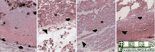
����ͼ4������Ĥɫ����Ƥ��Ӧ��(��b��)������Ĥɫ����Ƥ�벣��Ĥ�γɵļв��ڵ�����ϸ����������(Nc)������Ĥ����(��)��PAS��400��ͼ5������Ĥ��������(����)������Ĥ�ṹ���ƻ�(��)������ϸ��(RB)λ������ĤëϸѪ�ܲ�(��ͷ)���桡PAS��400��ͼ6������Ĥ��������(����)������ϸ��(RB)��������ĤëϸѪ�ܲ���(��)������Ѫ�ܲ�(��ͷ)����HE��200��ͼ7������Ĥ��������(����)������ϸ��������Χ��������Ĥ(��)����Ĥ��ͬʱ����(��ͷ)��HE��200��Fig.4��Reactive phase of retinal pigment epithelium(phase ��b).The tumor cells that were located among retinal��pigment epithelium and Bruch��s membrane had necrosis (Nc).The bruch��s membrane was intact(arrow)��PAS��400��Fig.5��Early phase of choroidal invasion(phase ��).The structure of Bruch��s membrane was destroyed(arrow).The tumor cells(RB)were localed at the surface of choroidal capillary layer(arrowhead)��PAS����400��Fig.6��Middle phase of choroidal invasion(phase ��).The tumor cells(RB)infiltrated into choroidal capillary(arrow)and middle blood vessel layers(arrowhead)��HE��200��Fig.7��Late phase of choroidal invasion(phase ��).The tumor cells infiltrated deep into the choroid(arrow)and involvement of sclera(arrowhead)��HE��200
����3������
����RB����ժ����������֯����ѧ�����ж�RB����Ԥ������Ҫ����ˣ��ڳ���Ĺ⾵��֯ѧ����У�������ϸ��������֯�ڵĽ���̶�����һ����֯ѧ�ϵ��жϱ���������ϸ��������Ĥ������Ԥ��Ĺ�ϵ��˵��硣���Ҫ��һ������Ͽɵ���֯ѧ����ͳһ��ʶ�������ƶ���ɫ����Ƥ-����Ĥ���ڿ��ǵ���RPE��BruchĤ������ϸ��������Ĥ��������е���������(����)��9�ݣ���������ϸ����������Ĥ�ķ�Χ�����С�̶Ƚ��л���(���ڣ�����)���ڹ⾵���ױ��ϣ������ڲ�ͬ�ĸ�����о��ж�����ϸ���Ľ���̶Ƚ��бȽϡ�
��������ϸ������Ĥ����ı����Լ���Ԥ���Ӱ����ұ�����һ��һ�㳣�没����Ƭ������Ĥ����ļ����Ϊ22��0%��43.2%��3-6�ݣ���������Ƭ�ļ���ʿɴ�62��0%��4�ݡ���Щ����ǽ����ڲ�ͬ������Ĥ������ڵĻ����ϵģ�������Խ��бȽϡ��жϡ��������ǵķ��ڵó����黼������Ĥ����ı���Ϊ24.24%����Shields�ȣ�3�ݵĽ��(23��0%)һ�¡�����(����Ĥȫ�����)����������ķ����ʽϸ�(22/26��84.6%)������ɸ�������34.6%��ת���ʸߴ�12��0%����0�����ڵ�30����Olver�ȣ�6�ݵ��о��������������Ĥ����ĸ߷����ʶ�Ԥ����Ҫ����Ҫ��������Ĥ����ķ�Χ��С��Shields�ȣ�3������Ϊ������Ĥ������Ԥʾת�Ƶ���Ҫָ���������ǵ�ͬʱ����������ʱ������������������ƣ���Ԥ��ĸ��ƿ���һ�������á�����26�����ڻ�����20�����������ƣ�����2�������18������ˣ��Բ�����Ϣ��ڵĻ��ߣ�Ҫ���������������ͬʱ�����������ߣ��䷢��ת�ƵĿ������������ߣ��б�Ҫ����������(����贩��)��������Ӧ�Ļ��ƻ�������ơ�
����4�������
����[1]��Heinrich TH,Messmer EP,Hoepping W,et al.Das metastasierun-
����gsrisiko beim retinoblastom.Klin Mbl Augenheilk,1991,199:319-324.
����[2]��Acquaviva A,Capolongo��A,Hadjistilianou TH,et al.Risk and influence factors for metastasis as well as for second non-ocular tumors in retinoblastoma patients.In��Bornfeld N��ed.Tumors of the eye��proceeding of the international symposium on tumors of the eye.Amsterdam:Kugler Publications��1991��145-147.
����[3]��Shields CL,Shields JA,Baez KA,et al.Choroidal invasion of��retino-
����blastoma:metastatic potential and clinical risk factors.Br J��Ophth-
����almol,1993,77:544-548.
����[4]��Redler LD,Ellsworth RM.Prognostic importance of choroidal invasion in retinoblastoma.Arch Ophthalmol,1973,90:294-296.
����[5]��Stannard C,Lipper S,Sealy R,et al.Retinoblastoma:correlation of invasion of the optic nerve and choroid with prognosis and metastasis.Br J ophthalmol,1979,63:560-570.
����[6]��Olver JM,McCartney ACE,Kingston J,et al.Histological indicators of the prognosis for survival following enucleation for retinoblastoma.In��Bornfeld N��ed.Tumors of the eye��proceeding of the international symposium on tumors of the eye.Amsterdam��Kugler Publications,1991��59-67.
����[7]��Zimmerman LE.Retinoblastoma and retinocytoma.In��Spencer WH��ed��Ophthalmic pathology��an atlas and textbook.vol.��.3rd ed.Philadelphia:W��B��Saunders,1985��1292-1351.
����[8]��Murphree AL,Rother C.Retinoblstoma.In��Ryan SJ��ed��Retina.vol��1St.Louis��Mosby,1989��517-556.
����[9]����죬Schilling H,Effert R,et al.297������Ĥĸϸ��������ɫ����Ƥ�仯��ͬ��ҽ�ƴ�ѧѧ����1998��27��103-105�� ��һҳ [1] [2] |