摘要 目的 观察各类滴眼剂对人角膜上皮细胞的毒性,以便提供临床上对药物的评估。方法 应用组织培养的方法,观察30种滴眼剂的毒性。结果 氯霉素(pH6.7)、庆大霉素(pH5.1)、磺胺类、喹诺酮类、咪唑类药物、地卡因、氧氰化汞等毒性较大。皮质类固醇的晚期毒性明显。结论 我们认为滴眼剂的毒性与主药、防腐剂和溶剂的毒性有关,特别是pH值。为此我们要注意配方的科学性,妥善的存放和合理的用药。
Toxicity of various kinds of eye drops to corneal
epithelium—a tissue culture assay study
ZHANG Luo,QIU Liang-Xiu,SHEN Yuan-Ping
Abstract Objective To represent an evaluation of drugs in clinics by investigating the toxicity of various kinds of eye drops to human corneal epithelium.Methods Tissue culture assay was applied to investigate the toxicity of thirty kinds of eye drops in vitro.Result The toxicity of chloramphenicol(pH6.7),gentamicin(pH5.1),sulfonamides,fluoroquinolones,dicaine and hydroarygli oxcyanide were serious.Corticosteroids were more toxicant in late stages.Conclusion The toxicity of eye drops are related to several factors:the main drugs,the preservative agents,the disolved solution,particularly the pH values.
Key words eye drop;toxicity;corneal epithelium
眼科局部药物治疗是治疗学上一个重要途径,而滴眼剂是常用的剂型。在临床工作中,我们常常遇到滴药后眼部有刺疼,或引起球结膜充血,更严重时发生角膜点状荧光素钠着色。为了进一步了解滴眼剂对角膜上皮细胞的毒性。我们采用组织培养的人角膜上皮细胞,作体外药物毒性试验,观察常用30种滴眼剂的毒性,为临床上选用、配制和使用滴眼剂提供参考。
1 材料和方法
1.1 人角膜上皮细胞 由我科实验室提供,用1640或Eagle培养液,加入100mL·L-1小牛血清,3g·L-1谷氨酰胺,20×103U·L-1庆大霉素,pH7.2。细胞量150×106个·L-1。置入24孔塑料培养板中,用烛缸CO2培养法,在37℃恒温箱内培养36~48h,此时细胞已贴壁并融合生长,待用,见图1。
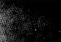
Figure 1 The tissue culture human corneal epithelial cells in vitro.spindle shapes attaching wall growth.contrast microscopy×100
组织培养人角膜上皮细胞倒置显微镜下,可见梭形贴壁生长正常
1.2 滴眼剂 庆大霉素、氟哌酸、抗自由基和抗胶原酶药物由我科实验室配制。计有抗生素、磺胺类、喹诺酮类、抗真菌类、皮质类固醇类、抗自由基和抗胶原酶等药物,共30种。
1.3 毒性实验 将备用的人角膜上皮细胞的培养液吸去,加入各类滴眼剂0.5mL,每种分4孔,加药后分别在15、30、60和120min用倒置显微镜进行观察,急性细胞毒性变化标准分3级[1]。加药后2h,将药液吸去,用生理盐水洗净,再加入培养液,在温箱内再培养24h,然后用台盼蓝染色,在显微镜下观察,计算死亡细胞数,以确定晚期细胞毒性,分3级[1],见图2~5。
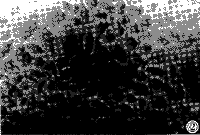
Figure 2 Corneal epithelial cells.Toxicity I grade "+" cytoplasm cloudy,small
particles here,contrast microscopy×250
角膜上皮细胞“+”毒性反应,胞浆透明度下降,出现小颗粒,倒置显微镜×250。
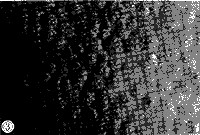
Figure 3 Corneal epithelial cells.Toxicity Ⅱ grade "+" particles in cytoplasm and nucleus,contrast microscopy ×250
角膜上皮细胞“++”毒性反应,胞浆和胞核出现颗粒,倒置显微镜×250。
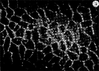
Figure 4 Corneal epithelial cells.Toxicity Ⅲ grade "+++" cell shrinked,pericellular widen,contrast microscopy×250
角膜上皮细胞“+++”毒性反应,可见细胞皱缩、间隙加宽,倒置显微镜×250。
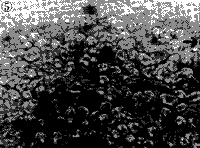
Figure 5 Corneal epithelial cells.Toxicity in late stage"+" ,Typan blue staining×250 角膜上皮细胞晚期毒性“+”,“↑”台盼蓝着色细胞×250
[1] [2] [3] 下一页 |
