摘要 目的 检测增生性玻璃体视网膜病变(PVR)发生过程中白细胞介素-1β(IL-1β)的含量变化。方法 用同种巨噬细胞诱发兔眼PVR,在不同时间点抽取玻璃体液,用双抗体夹心法酶联免疫检测(ELISA)试剂盒测定IL-1β含量。结果 IL-1β含量在7天时明显增加,在14~28天间呈一较高水平(175~171ng/L);21天达高峰值234ng/L,其基线含量为4ng/L。结论 IL-1β在此模型的含量在7~28天明显增高,与PVR病程的炎症期、增生期和瘢痕期相吻合,提示在PVR发生中对细胞增生和瘢痕化有调控作用。
IL-1β in experimental proliferative vitreoretinopathy induced by macrophages
Shi Yining Hui Yannian Yang Kun
(Department of Ophthalmology,the Fourth Hospital of Xi’an,Xi’an
710004)
Abstract ObjectiveTo investigate the concentration changes of interleukin 1β(IL-1β) in the the development of experimental proliferative vitreoretinopathy(PVR)induced by macrophages.MethodsThe cell-cultured macrophage suspension was injected into the rabbits intravitreally for PVR inducement.The animals were divided into 4 groups,and were observed on 7,14,21,and28d,respectively.The criterion of this PVR model is 0,1~4 stages.The vitreous were extracted pre-and post-injection at the above point of times.IL-1β levels in them were measured with an enzyme linked immunosorbent assay (ELISA) kit.The data were calculated statistically.Results The PVR formation corresponded to the previous result.The trend of concentration changes in the PVR vitreous of IL-1β went up from 0d to 21d,and reached its peak 234ng/L at d 21,which was parallel to the inflammatory and cellular proliferative stages of PVR,then IL-1β kept the high level in the scarring stage and maintained a plateau between d 14 and d 28(175~171ng/L).There were no significant changes of the concentrations in vitreous and serum of the control eyes during the experiment.Conclusion Since the macrophages were the only cells induced into the rabbits’ vitreous cavity in this PVR model,all proliferative cells were derived from the hosts by themselves.And the inflammatory stage was the starting of the PVR model which may help to reveal the changes and the mediating role during the PVR formation.The increasing of IL-1β in the vitreous parallels to the inflammatory and proliferative stages and then maintained its plateau at the regenerative stage which means it may play an important role in initiating and mediating the cellular proliferation and post-inflammatory regeneration in the vitreous.The data show the secretion type of cytokines in the eye belongs to the autocrine and paracrine without nervous and indocrine-like regulation.
Key words proliferative vitreoretinopathy macrophage interleukin-1β vitreous enzyme linked immunosorbent assay
增生性玻璃体视网膜病变(proliferative vitreoretinopathy,PVR)是一种眼内组织损伤后过度修复反应,同时伴有复杂的免疫调节机制[1]。损伤初期的炎症是启动细胞增生修复的先决条件[2]。以往人们对此认识不足,研究较少。我们采用巨噬细胞诱发的炎症性PVR模型,对炎源性细胞因子之一的白细胞介素-lβ(interleukin 1β,IL -1β)在病程中的动态变化进行了定量检测。
1 材料与方法
1.1 巨噬细胞的采集与纯化 方法同以往报告[3]。将上述细胞悬液注入玻璃体腔视盘前方,100μl/眼;模型眼PVR诱发结果的判定标准同文献[4]。每只兔的对侧眼为空白或单次抽取玻璃体液后的对照眼。
1.2 标本的收集和贮存 (1)兔眼玻璃体液采集:分别于注入巨噬细胞前即刻,以及注入后7、14、21、28天5个时间点分别对16只兔32只眼自鼻上角膜缘后2mm抽取玻璃体液100μl/眼,冷存待测。每只兔仅抽取1次,随后处死动物,摘除眼球,立即固定于10%福尔马林液中,作常规病理检查。(2)兔静脉血采集:每组兔处死前抽静脉血2ml,6000r/min离心5min,抽吸血清100μl,贮存。
1.3 IL-1β检测 (1)液体样品:采用深圳晶美生物技术有限公司提供的双抗夹心法ELISA检测IL-1β试剂盒。标准品倍比稀释10个浓度,设零孔、空白孔为对照。每孔加样品100μl。具体操作见试剂盒说明书。显色后在450nm处ELIAS读数仪测A值。绘制标准曲线,计算标本中IL-1β浓度。(2)组织标本:对兔眼视网膜组织行间接免疫荧光组织化学检测。第一、二抗体由第四军医大学免疫学教研室和病理学教研室提供,方法同前文报告[5]。
1.4 统计学处理 采用SPLM、EUS2.0、SAS6.04及HG2.3统计软件对实验数据进行t检验、多元方差及多元回归拟合曲线分析。
2 结果
2.1 玻璃体内IL-1β含量 IL-1β的玻璃体内含量在7天时明显增加(95ng/L),至21天达峰值234ng/L,28天趋于下降(171ng/L),在14~28天间呈一较高水平(175ng/L~171ng/L)。统计学处理有显著性差异(表1,P=0.0006)。
表1 巨噬细胞诱发的兔PVR眼玻璃体内IL-1β含量变化( ±s) ±s)
Tab.1 The changes of concentration of IL-1β in the rabbit vitreous of
the macrophage induced PVR( ±s) ±s)
| |
Time(d) |
| 0 |
7 |
14 |
21 |
28 |
P1 |
| Model eyes(ng/L) |
5±2 |
95±142 |
175±130 |
234±304 |
171±192 |
0.4685 |
| Control(ng/L) |
3±2 |
2±1 |
3±3 |
4±1 |
2±1 |
0.6729 |
| P2 |
0.2544 |
0.2390 |
0.0382 |
0.1818 |
0.1290 |
|
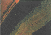
P1:t test within group;P2:mono cariate analysis between groups;2.2 视网膜组织内IL-1β水平 (1)模型眼PVR的判定采用文献中的0、Ⅰ~Ⅳ5个级别,7天组Ⅰ级1眼,14天组Ⅰ级、Ⅱ级各1眼,21天组Ⅰ级2眼、Ⅲ级和Ⅳ级各1眼,28天组Ⅲ级3眼、Ⅳ级1眼,均无玻璃体出血;(2)PVR模型眼经抗IL-1β单抗的间接免疫荧光染色,1~2周可见视网膜色素上皮细胞(retinal pigment epithelium,RPE)胞浆内有较弱荧光染色;3~4周RPE至视锥层外2/3及内网状层内呈强荧光染色。玻璃体前膜内亦呈高荧光(图1)。

图1 PVR模型眼28天视网膜组织IL-1β的间接免疫荧光染色,×40
Fig.1 Indirect fluorecein staining of FITC-labeled IL-1β
in the retina of PVR model at 28d.FITC×40
2.3 玻璃体内IL-1β基线水平 零天空白对照眼玻璃体内IL-1β含量为(3±2)ng/L;眼内注入巨噬细胞及单次抽取玻璃体液刺激后,玻璃体内IL-1β含量在各时间点的波动无显著差异(P=0.2045)。
2.4 血清内IL-1β基线水平 正常兔血清内IL-1β含量(95±107)ng/L;眼内注入巨噬细胞后,血清内IL-1β含量无明显变化(P=0.5005)。
[1] [2] 下一页 |
