摘要 目的 探讨副泪腺的分泌 机制,提供副泪腺神经支配的形态学证据。方法 10例人眼标本,男性9人,女性1人,年 龄20 ~38岁,用标准透射电镜技术对42个Krause腺Wolfring腺进行观察。结果 副泪腺间质中广泛存在大量无髓神经纤维,轴突终末与肌上皮细胞形成突触结构,突触间 隙 10~30nm;裸露的轴突终末穿过腺上皮基膜,走行于腺上皮细胞间并与之形成突触结构 。轴突终末膨体内含有大量圆形清亮突触小泡及少量中央为致密核芯的大颗粒小泡,是典型 的副交感神经纤维。结论 人类副泪腺存在神经支配,其性质以副交感为主并有多种神经 递质参与。
Are accessory l acrimal glands innervated?
——The fine structure of nerve terminations in the human accesso ry lacrimal glands
Cui Hongping Qiu Xia ozhi Yu Zhang
(Department of Ophtha lmology,Eye and ENT Hospital of
Shangha i Medical University,Shanghai 20003)
AbstractObjectiveTo study the secretion m echanism of human accessory lacrimal gla nds and to document morphologic evidence for the functional innervation of acces sory lacrimal glands.Methods42 access or y lacrimal glands were excised from 10 e yes enucleations (aged 20~38,male 9,fema le 1),fixed with 2.5% glutaraldehyde in 0 .1 mol/L phosphate buffer (pH7.4),post-f i xed in 1% osmic acid,and embedded in Epo n618.Stained with uranyl acetate and le ad citrate,ultrathin sections from each accessory lacrimal gland were examined w ith TEM-1200EX electron microscope.Res ultsNon-myelinated nerve fibers in gre at number were found in the stroma of acce ssory lacrimal glands.Axons lay either deep within Schwann cells or ran along t he periphery of Schwann cells.The diamet e rs of axon varied 200nm~1μm.The axopla smic organelles included mitochondria ne urotubules and different kinds of vesicl es.Some of fine single naked axons invag inated into myoepithelial cells and were identified forming the synapses directl y with myoepithelial cells,while ot hers penetrated the basement membrane of acinar cells to pass in between them.Th e distance between axons and target cell s ranged from 10~30nm.In the varicositi es of these axon terminals there were ma ny small clear vesicles,with diameters 2 0~50nm,and a few larger granular vesicl es,with diameters 80~100nm.The small cl ear vesicles are considered containing a cetylcholine (ACh) mainly while large gr anular vesicles containing neuropeptides .So,those axon terminals were considered typical of cholinergic parasympathetic nerve fibers morphologically.Conclusion The results indicate that human access or y lacrimal glands are definitely innerva ted.The nerve fibers of functional inner vation are parasympathetic structures mo rphologically.In addition to ACh,several kinds of neuropeptides may also act as neurotransmitter.
Key words accessory lacrim al gland synapse non-myelinated nerve fi ber parasympathetic nerve
传统观点认为,副泪腺是没有神经支配的外分泌腺,它主要分泌基础泪液中的水性成分,而主泪腺是有神经支配的,独立承担反射性和心因性泪液分泌[1]。 许多学者通过间接的证据,对副泪腺无神经支配这一观点提出了质疑[2~5],但未见有力的形态学依据。直到1994年Seifert等[6]才首次发表了副泪腺存在神经支配的电镜照片,但迄今尚缺乏进一步深入研究,为此,我们对人副泪腺中的神经末梢进行了全面的电镜观察。
1 材料与方法
全部材料取自10例脑外伤死亡者,年龄20 ~38岁,男性9人,女性1人。在解剖镜下迅速取包含有krause腺的穹窿部结膜组织和包含有Walfring腺的睑板上缘组织,仔细剖切成1mm×1mm×1mm大小的组织块,用2.5%戊二醛固定,其后步骤按标准透射电镜程序进行,用日本电子公司的 TEM1200EX透射电子显微镜进行观察。
2 结果
2.1 副泪腺间质中的神经末梢
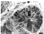
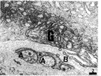
在副泪腺组织间质中观察到许多粗细不等的无髓神经纤维(图1 ~2)。不同数量的轴突全部或部分包埋在雪旺细胞表面凹陷形 成的纵沟内。轴突的直径从200nm~1μm,轴质(axoplasm)中可见线粒体、微管、神经丝和包含有神经递质的小泡。小泡主要有两种,直径20~50nm的圆形清 亮突触小泡和直径80~100nm的大颗粒小泡。颗粒小泡的中央为致密核芯,核芯的周围是透明。

图1 透射电镜下所见的副泪腺组织(G)形态,可见组织间质的
神经末梢(箭头)。×1000.Bar 5 μm
Fig.1 Glandul ar tissue of accessory lacrima l gland.Arrow
shows nerve fiber;G:glandu lar epithelial cells.×1000.Bar 5μm

图2 副泪腺组织间质中的无髓神经纤维。轴突(A),基膜(B),
雪旺细胞(S),腺上皮(G).×20K.Bar 200n m
Fig.2 Non-myeli nated nerve fiber in connective tissue o f an accessory
lacrimal gland.Axon(A),Ba sement membrane(B),Schwann cell(S),
Gland ular epithelial cells (G).×20K.Bar 200n m
2.2 神经末梢与肌上皮细胞的连接
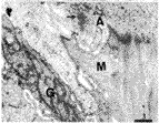
轴突终末与肌上皮细胞形成“突触结构”(图3~4)。轴突终末周围无雪旺细胞的胞膜而呈完全裸露,位于肌上皮细胞胞膜凹陷形成的浅槽内(图3)或与肌上皮细胞胞膜平行(图4),二者之间的突触间隙10~30nm,在突触前膜和突触后膜未见明显的致密带,不参与形成突触的轴膜表面覆以基膜。突触前轴突终末膨体(varicosity)内可见线粒体、大量圆形清亮突触小泡和少量大颗粒小泡。在图3中还见到3个中央的致密核芯明显较小的颗粒小泡。肌上皮细胞以胞浆中含有大量肌动蛋白丝为特征,其与腺上皮关系密切,图4中可见其与腺上皮之间形成的桥粒连接。

3 轴突终末(A)与肌上皮细胞(M)形成的突触结构。轴突终末膨体内含小圆形清亮突触
小泡(箭头)和大颗粒小泡(三角符),是典型的副交感神经纤维。×12K.Bar 500nm
Fig.3 Synapse between axons terminal (A) and myoepithelial cells(M).
The varicosity contains small clear vesi cles (arrow) and larger granular
vesicle s (arrow head),typical of cholinergic pa r asympathetic
innervation.×12K.Bar 500nm

4 轴突 终末(A)与肌上皮细胞(M)形成的突触结构。可见肌上皮细胞与腺上皮细胞间的
桥粒连接(箭头)。×12K.Bar 500nm
Fig.4 A nother type of synapse between axons ter minal (A) and
myoepithelial cells(M).Des mosome between myoepithelial cell and gl
andular epithelial cell (arrow).×12K.Bar 500nm
2.3 神经末梢与腺上皮细胞的连接
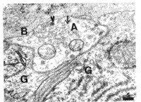
完全裸露 的轴突终末穿过腺上皮的基膜而走行于副泪腺腺细胞之间(图5),并与腺细胞形成突触结构,二者间的间隙10~30nm。轴突终末膨体内同样含有线粒体、大量圆形清亮突触小泡和少量大颗粒小泡。

5 位于腺上皮(G)之间的单一裸露的轴突终末(A ),内含小圆形清亮突触小泡(箭头)和大颗粒小泡(三角符)。×12K.Bar500 nm Fig.5 Naked s ingle axon terminal(A) with sma ll clear vesicles (arrow) and larger gra nular vesicles (triangle) between glan dular epithelial cells (G)and basement membrane(B).×12K.Bar 500nm
[1] [2] 下一页 |
