����ժҪ��Ŀ�� �о������Ĥ��ֲ�ųⷴӦ���� ���������� ����ģ��(Wistar ����18ֻ������9ֻ)���۲��Ĥ���ȡ�����Ѫ�ܡ���Ĥ������Ѫ������⡣���Խ�Ĥ�� ��ɨ��羵�۲졣��� ����1�ܣ�ֲ����������Ѫ�ܡ���Ĥˮ�ס�������������1�£� ֲ���ۻ�����ˮ���ᡣ�����黯��ʾ����ֲ��3�죬Ѫ��Th��iϸ��IL-2R��ϸ�����ߣ�15��ʱ��ĤֲƬ������TC��Sϸ������Ϊ�����ܰ�ϸ�������� �����Խ� Ĥ��ֲ�����ųⷴӦ��������������������Ƶ���ֲ�ų���̡�
The cl inical immunology study of cor neal penetrating xenograft in Wistar rats
Ding Wei��Wu Guijun��Chen Jiaqi
������Department of Ophthalmology��Nanfang Hospi tal��First Military Medical
����University��Gu angzhou 510515��
����Abstract ObjectiveTo in vestigate the rejection mechanisms of co rneal penetrating xenograft��MethodsTh e corneal penetrating xenograft models wer e made in 18 Wistar rats and9 guinea pi g s��The diameter of graft was 6.0mm��and diameter of receipt bed was 0.5 mm shorter than the graft��The graft was continuous sutured by nylon wire��Cornea l pellucidity��edema and neovascularizati on were observed by opton slit-lamp micr oscopy every 4d��and stopped till 30d a fter keratoplasty��Rejection value ��RV�� w as calculated according to Holland��s gra de��On 6��10d ��one eyeball��and 15d��two e yeballs�� after keratoplasty��immunohistoc hemical study was performed in the corne a��On 3d preoperation and 3��9��15��21��30d postoperation��1.5mm blood was gotten o ut from the rat��s hearts��and the change s of lymphocyte subgroup were observed��O n 12��20d after keratoplasty��the corneal constitution and form of the animals we re respectively observed and taken a pho tograph by scanning electron microscope�� I mmunological study was performed in the cornea and the peripheral blood��Results In the first week of postoperation��neo va scularization��corneal edema and thicknes s of stroma occurred in recipient site��I n the first postoperative month��the reci pient site cicatrized and corneal edema lessened��Immunohistochemical studies sho wed that Th��i cells and IL-2 R�� cells w ere increased in the peripheral blood on 3d after keratoplasty��At 15d after ke ratoplasty��the major infiltrating cells were Tc��s cells in the graft corneas��Co nclusionThe rejection of corneal penet ra ting xenograft in rats may be similar to that of other organs��
����Key words��cornea l penetrating xenograft immunological study
���������˺�Ⱦ�Խ�Ĥ�������ɵ��½�Ĥ���ǣ������½���Ŀǰ�����Խ�Ĥ��ֲ�Թ���Ϊ���������Խ�Ĥ���ǡ��ָ�������Ψһ��Ч���������������ųⷴӦ����в��Ĥ��ֲƬ�ɻ��������ʧ�ܵ�Ҫ��֮һ�����ִ��Խ�Ĥ��ֲ�����ųⷴӦ������ˡ�Ϊ̽�����ִ��Խ�Ĥ��ֲ�����ųⷴӦ�ķ������������������Wistar��������������ִ��Խ�Ĥ��ֲ���۲�������ѧ�仯������̽���������ִ��Խ�Ĥ��ֲ�����ųⷴӦ�Ŀ��ܷ���������
����1�������뷽��
����1��1��Wistar����(2��3���䣬����180��220g������)18ֻ������(2��3���䣬����200��230g�����ۼ���)9ֻ��
���������黯��Ҫ�Լ���С����T�ܰ�ϸ����ȺW��OXϵ�е���¡���壬�ɾ���ҽѧ��ѧԺ���ڷ�װ����ʾϵͳ��ABC�Լ��У�ΪDako��˾��Ʒ��
����1��2���������ִ��Խ�Ĥ��ֲ��(��Ϊ���ۣ���������)
����1��3���ٴ��۲�
��������Holland����6���������ٴ��۲����ַ����۲��Ĥ���ǵ÷�ֵRV��rejection value��Ĥ���Ƿ�ֵ)���Ʒֱ����£�
�������ȣ�(0)������(1)��Ȼ��ǣ�(2)���Ǽ��أ�ǰ������ɼ���(3)���Ǽ��أ�ǰ��ģ���ɼ���(4)ȫ���ǣ�ǰ����������
����ˮ�ף�(0)����ˮ����Ƥˮ�ף�(1)��Ȼ�������(2)�����Ի���ˮ�ף�(3)�����Ի���ˮ�ײ���ƤMicrocyticˮ�ף�(4)�����Խ�Ĥ���䡣
��������Ѫ�ܣ�(0)�ޣ�(1)�ܱ�����Ѫ�ܣ�(2)�пڲ�����Ѫ�ܣ�(3)ֲƬ����Ѫ�ܣ�(4)ȫ��Ĥ����Ѫ�ܡ���Opton��϶�������۲죬ÿ��3��Ʒ�1�Σ���̬�۲�30�졣
����1��4�����߲����۲�
������Ĥ�����黯����Ĥ��ֲ�����6��10���ȡ1ֻ����15��ȡ2ֻ���Ĥ��ֲ������������Ƭ�������黯��
���������ܰ�ϸ����Ⱥ��̬��������ǰ3�죬����3��9��15��21��30�죬���ڴ���ȡѪ1.5ml���۲��ܰ�ϸ����Ⱥ�ı仯��
����1��5��ɨ��羵����Ĥ��ֲ�����12��20���ȡ1ֻ��Ĥ��ɨ��羵�£��۲�ֲƬ��ֲ����֯ϸ���Ľṹ����̬�仯����Ӱ��
����2�����
����2��1���ٴ��۲�
��������Holland�Ʒַ����۲��ųⷴӦ�̶ȵĶ�̬����(��ͼ1)������1�ܣ�ֲ����������Ѫ�ܣ���Ĥ���Ǽ��أ���������ˮ�ף�15��RV�ӽ���ֵ��1��ʱ��ֲƬ�����ۻ���ֲ��ˮ���ᣬһ������ʱֲƬ�ۻ���������ֲ��ˮ�����ˡ��������Ĥ��ֲ�����Ĥһ����ˮ�ף�1�ܺ��ĤֲƬ����
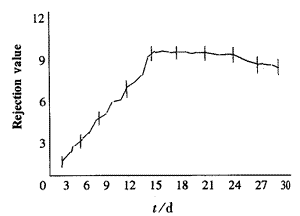
���� ͼ1����Ĥ��ֲ���ųⷴӦ�̶ȵı仯
����Fig��1��The changes of reject ion value after corneal graft
����2��2����ֲǰ��Ѫ���ܰ�ϸ����Ⱥ�ı仯
������Ĥ��ֲǰ������ѪTϸ����Ⱥ�Ķ�̬�۲����1��
��1����Ĥ ��ֲǰ������ѪTϸ����Ⱥ�Ķ�̬�۲�( ��s) ��s)
����Tab��1��Changes of the T cells in per ipheral blood before corneal graft(��s)
|
Time��d��������n |
Th��i |
IL-2R�� |
Tc |
|
Preoperative 3 ��8 |
3.3��1.94 |
2.3��0.6 |
23.1��2.4 |
|
Postoperative �� 8 |
|
3 |
47.2��1.2 |
5.1��0.5 |
24.8��2.7 |
|
9 |
46.0��1.4 |
6.8��0.7 |
30.4��2.4 |
|
15 |
47.1��1.1 |
7.9��0.7 |
34.7��3.5 |
|
21 |
46.1��1.7 |
7.6��0.8 |
36.3��1.2 |
|
30 |
44.3��2.8 |
8.1��0.3 |
32.8��1.6 |
�����ɱ��пɼ�������3�죬Th��i��IL-2R��ϸ���ٷ��ʿ�ʼ�������ߣ�����9�죬Th��i���н��ͣ�IL-2R��ϸ���仯����Tc��s�������ߡ�
����2��3�������ĤֲƬ�ܰ�ϸ����Ⱥ�۲�
�����ӽ�Ĥ�����黯�ɹ۲쵽������6�죬ֲƬ�г���Ia��ϸ����������10�죬ֲƬ�ܰ�ϸ��������أ����Է��ߴ�Ϊ������Ia��ϸ�����ࡣ����15�죬ֲƬ�д������ܰ�ϸ������Tc��s����ϸ���϶࣬Th��i��IL-2R��Ia���б��
����2��4��ɨ��羵�۲�
��������12�죬ֲƬ�п��ڼ���Χ���ܰ�ϸ��������T�ܰ�ϸ��������ϸ�������ֲƬ��Ƥϸ����϶������ë���١�����20�죬ֲƬ��Ƥ�ų⣬ϸ��Ĥ���ѡ�������
[1] [2] ��һҳ |