摘要 目的 探讨细胞间粘附分子-1(ICAM-1)在角膜基质细胞中 的表达,以及炎症介质对其表达的调节作用。方法 采用细胞培养、免疫细胞化学和流式 细胞技术,观察人角膜基质细胞ICAM-1的表达及脂多糖(LPS)和干扰素-γ(IFN-γ)对此表 达的影响。结果 体外培养的基质细胞可基础表达一定量的ICAM-1,LPS和IF N-γ可上调其表达水平至基础表达的1.4~4.6倍。结论 角膜炎症反应中,炎症介 质促进基质细胞表达ICAM-1,与白细胞表面的配体结合导致白细胞的迁移、聚集和局 部炎症反应的产生。
Expression and modulation of inte rcellular adhesion molecules-1 on cultu red
human corneal keratocytes
Li Haili Wu Jingan Sun Xi ngcai
(Department of Ophthalmology,the Fi rst Teaching Hospital,Beijing Medical
U niversity,Beijing 100034)
Abstract Object iveTo investigate the expression of in tercellular adhesion molecules-1(ICAM-1) and its regulation by inflammatory fact ors in human corneal keratocytes.Method sHuman corneal keratocytes were establ ish ed in 10-d to 30-year-old donor eyes.T he cubes of corneal stroma were cultured in media consisting of DMEM supplemented with 20% fetal calf serum.The second pa ssage cells were seeded at high density, incubated to subconfluent,and activated with lipopolysaccaride (LPS) and interfe ron-γ(IFN-γ).The constitutive expression of IC AM-1 by the third passage human corneal keratocytes was assessed by immunocytoch emical staining and flow cytometry analy sis.In addition,the regulation of the ex pression of ICAM-1 by LPS and IFN-γ was determined.Results The behavioral char ac teristics and morphological features of third passage cultured human corneal ker atocytes were similar to those in vivo.T hese cells were suitable for experiments of corneal diseases in vitro.Immunohist o chemical staining demonstrated that cult ured human corneal keratocytes can const itutively express certain amount of ICAM -1.After exposure 24h to either LPS or IFN-γ,progressive increased significantl y in ICAM-1 staining occurred.Flow cytom etry analysis determined that ICAM-1 was constitutively expressed by cultured co rneal keratocytes.After LPS and IFN-γ st imulation for 12 and 48h respectively,t here was a median fold increase in expre ssion of 1.4~4.6.The upregulation of LPS and IFN-γ on ICAM-1 by keratocytes was related to the stimulating period and co ncentration of each factor.Conclusions I n corneal inflammation,inflammatory fact ors can enhance the expression of ICAM-1 in keratocytes.Its binding with the lig and on the surface of leukocytes leads t o migration and aggregation of leukocyte s in inflammed cornea and thus results i n local inflammation.Key wordsinterce llular adhesion molecules-1 human cornea l
keratocytes lipopolysaccaride interfer on-γ
细胞间粘附分子-1(intercellular adhesion molecule-1,ICAM- 1)是参与炎症反应的重要细胞粘附分子之一。已有研究表明,炎症角膜组织中基质细胞表达ICAM-1增加,基质细胞是炎症反应的主要靶细胞 。因此,研究炎症介质对体外培养的人角膜基质细胞表达ICAM-1的调节,对于阐明角 膜炎症反应的发生机制有重要意义。
1 材料与方法
1.1 实验材料
标本来源于北京医科大学第一医院眼库,出生1天至30岁的人眼球,外伤或急症死亡,24h内取材,离体后1h内接种。
主要试剂、抗体和仪器 (1)DMEM 培养液和0.25%Trypsin(GIBCO);胎牛血清(医科院血研所);0.05%DAB显 色液(中山公司);脂多糖(lipopolysaccaride,LPS)来自大肠杆菌( Sigma);人重组干扰素-γ(interferon-γ,IFN-γ)(Progm ega)。(2)鼠抗人ICAM-1单克隆抗体(santa);生物素偶联马抗小鼠IgG和 辣根过氧化物酶标记链霉亲和素(vector);FITC标记羊抗小鼠IgG(Jackson)。(3)流式细胞仪(FACstar,BD公司)。
1.2 实验 方法
1.2.1 取材及细胞培养 无菌条件下剪取1mm3大小的人角膜基质组织块接种于15cm2的培养瓶中,加入含20%胎牛血清的DMEM培 养液,于37℃5%CO2培养箱中培养。细胞汇合后,0.25%Trypsin消化,1∶2 或1∶3均匀传代。
1.2.2 免疫细胞化学(LSAB 法) 观察基质细胞ICAM-1的表达及其调节 同一标本传1或2代基质细胞均匀 传代至3组铺有盖玻片 的培养皿中,70%汇合时,分别加入含10μg/mlLPS、含500U/mlIFN -γ及不含药的无血清DMEM培养液,培养24h,冷丙酮固定;经抑制内源性过氧化物酶及 1%牛血清白蛋白(BSA)封闭,加鼠抗人ICAM-1单抗,4℃过夜;加生物素偶联马抗小鼠 IgGⅡ抗,370℃30min;加辣根过氧化物酶标记链霉亲和素,37℃ 40m in;0.05%DAB溶液显色。以PBS或BSA代替Ⅰ抗做阴性对照。
1.2.3 流式细胞仪检测LPS和IFN-γ对基质细胞表达ICAM-1的影响 (1)同一 标本传1或2代已达汇合的基质细胞均匀传代至数个35cm2培养瓶中,70%汇合时分别加入LPS或IFN-γ。分组:a)分别加入含5、10、20、30μg/ml LP S及不加药的无血清DMEM培养液作用12h。b)加入含LPS的无血清DMEM培养液后分别作 用6、12、24h(LPS浓度为a)中作用最强的浓度。c)分别加入含100、50 0、1000U/ml IFN-γ及不加药的无血清DMEM培养液作用48h。d)加入含500U/ml IFN-γ无血清DMEM培养液分别作用24、48、72 h。
(2)间 接免疫荧光标记及检测:a)制备单细胞悬液。b)计数细胞:每管≥1×10⁶个细胞,活细胞≥95%。c)加鼠抗人ICAM-1单抗,4℃50min,离心,弃上清。d )加FITC标记羊抗鼠IgG Ⅱ抗,4℃ 40min,离心弃上清,避光。e)荧光显 微 镜下观察,照相。f)流式细胞仪检测荧光阳性细胞数及阳性细胞平均荧光强度。测定免疫 荧光(FL1)时,每管计数5000个细胞,以对照管细胞测定标准基线。两对照管为: (a)PBS代替Ⅰ抗,(b)PBS代替Ⅰ抗和Ⅱ抗。
2 结果
2.1 免疫细胞化学染色:体外培养的人角膜基质细胞可基础表达一定量ICAM-1,主要位于细胞表面,高倍镜下呈细小的点状、颗粒状棕黄色着染。经LPS(10μg/ml) 和IFN-γ(500U/ml)作用后的细胞表达ICAM-1明显比未加药细胞增强,两加药组之间镜下所见差别不大。阴性对照不着染。
2.2 流式细 胞仪检测:间接免疫荧光标记后的基质细胞荧光染色阳性。以两对照管细胞测定相对荧光强 度基线(baseline)为11.25。
基质细胞可基础表达一定量的ICAM-1,荧光细胞阳性率为76.5%~94.1%,阳性细胞的相对平均荧光强度为30.66~71.54。经LPS或 IFN-γ作用后,阳性细胞数变化不明显,而相对平均荧光强度显著增加,达到基础表达 的1.4至4.6倍。
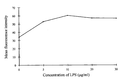
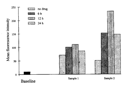
(1)LPS对基质细胞表达ICAM-1的影响:如图1所示,5μg/mlLPS即可上调基质细胞表达ICAM-1,10μg/mlLPS作用最 强,20μg/ml和30μg/ml的作用略有下降。选择10μg/ml为LPS作用浓度,观察LPS作用不同时间基质细胞表达ICAM-1的情况,结果为:LPS作用6h,基 质细胞表达ICAM-1明显增加,12h达高峰,最高达到用药前的4.6倍,24h明显下降(图2)。

1 角膜基质细胞表达ICAM-1与LPS浓度的关系
Fig.1 Relationship betwe en ICAM-1 expression of
keratocytes and the concent ration of LPS

图2 LPS(10μg/ml)作用时间对HCS表达ICAM-1的影响
Fig.2 Effect of LPS on ICAM-1 expression of keratocytes for various pe riods
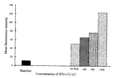
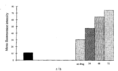
(2)IFN-γ对角膜基质细胞表达ICAM-1的影响:由图3和图4可看出,IFN-γ可上调角膜基质细胞表达ICAM-1,其上调作用呈浓度和时间依赖性。

3 不同浓度IFN-γ对HCS表达ICAM-1的影响
Fig.3 Effect of different concentration of IFN-γon ICAM-1 expression of keratocy tes

图4 IFN-γ(500U/ml)作用时间对HCS 表达ICAM-1的影响
Fig.4 Effect of IFN-γ (500U/ml) on ICAM-1 expression of
keratocytes for various periods
[1] [2] 下一页 |
