|
2 结果
油红染色阳性的组织切片中,Bruch膜呈红色。经脂溶剂处理后,Bruch膜染色呈阴性。年轻组中染色几乎均表现阴性(图1),仅一例28岁眼Bruch膜呈细线状轻度染色(图2);随年龄增长,Bruch膜的油红染色带加宽,染色增强(图3,4)。中年组,大多数眼呈轻度染色;老年组的染色均为中度及重度,Bruch膜呈均匀一致的带状染色,脉络膜的毛细血管间柱(intercapillary pillars)也有着色(图3)。RPE染色程度及面积,亦随着年龄增长而增加。20天,5个月及2岁龄眼中,黑色素颗粒遍布整个RPE细胞,而无染色(图1);RPE染色最初见一位8岁患者供眼,呈轻度,局限于细胞基底部;老年眼染色可占细胞总面积的2/3,且染色程度不受脂溶剂的影响,黑色素颗粒则逐渐减少至局限于细胞顶部(图3)。同一眼中,黄斑部Bruch膜及RPE细胞染色较周边部强。
AMD眼中,Bruch膜重度染色,玻璃疣及基底层状沉着(basal laminar deposits)均强烈染色;RPE细胞染色遍布细胞浆(图5)。
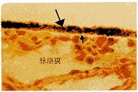
图1 2岁眼油红染色冰冻组织切片显微照片。Bruch膜(小箭)及RPE细胞(大箭)均无染色 ×400
Fig.1 Photomicrograph of the macular region section from a2-year-old donor.No staining of Bruchs membrane (small arrow)and RPE cells(large arrow)is demonstrated.Cryosections stained with oil red Oand counter stained with Mayers haematoxylin×400
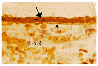
图2 28岁眼油红染色冰冻组织切片显微照片。Bruch膜(小箭)呈线形染色,RPE细胞(大箭)轻度染色 ×400
Fig.2 Photomicrograph of the macular region of section from a28-year-old donor.Light staining of Bruchs membrane (small arrow)in a thin line pattern and RPE cells(large arrow)is demonstrated.Cryosections stained with oil red O and counter stained with Mayers haematoxylin ×400
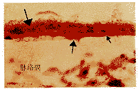
图3 72岁眼油红染色冰冻组织切片显微照片。Bruch膜(小箭)呈带状染色,毛细血管间柱亦有染色(中箭),RPE细胞(大箭)染色明显 ×400
Fig.3 Photomicrograph of the macular region of section from a72-year-old donor.Deep staining of Bruchs membrane(small arrow)in a broad line pattern and RPE cells(large arrow)is demonstrated.The choroid intercapillary pillar(middle arrow)is also stained.Cryosections stained with oil red O and counter stained with Mayers haematoxylin ×400
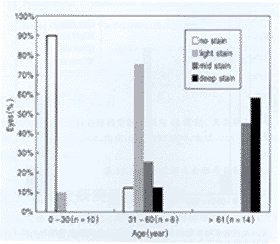
显示染色程度随年龄而增强,条形图代表各种染色程度在不同年龄组中所占的百分比(n=眼数)
图4 不同年龄组中Bruch膜油红染色程度示意图
Fig.4 The histogram demonstrates the different staining intensity of oil red O in Bruchs membrane of three age groups.The vertical bars represent the percentage of eyes with a particular staining intensity within each age group.The graph highlights an increasing staining intensity with age(n=number of donor eyes)
溴-苏丹黑 B阳性染色组织切片中,Bruch膜呈蓝黑色。经脂溶剂处理的组织切片,染色呈阴性。Bruch膜阳性染色表现及特点同油红。年轻组中只有1例(28岁)为轻度染色;中年组则多数为轻度染色;老年组中,64%表现为中度及重度染色(图6)。RPE细胞染色也可观察到,但因黑色素颗粒与此染色相近,不易区分。黄斑区与周边部Bruch膜及RPE细胞染色也有区别,但不如油红染色表现的清晰。AMD眼表现为溴-苏丹B的强烈染色,其染色特点与油红染色相似。
溴-丙酮-苏丹黑B阳性染色为灰色,其染色程度同上述两种脂类染色一样,随年龄的增长而增加。年轻组则均为阴性;中年组12.5%轻度染色;老年组50%呈轻度或中度染色(图7)。染色阳性占溴-苏丹黑B染色眼的40%。两例AMD眼中,只有1例中度染色。
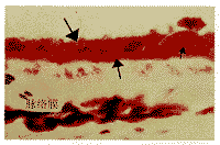
图5 AMD眼油红染色冰冻组织切片显微照片。Bruch膜(小箭)及RPE细胞(大箭)染色较正常老年眼更为强烈,玻璃疣drusen亦有染色(中箭) ×400
Fig.5 Photomicrograph of the macular region sections of an age-related macular degeneration eye from a 67-year-old donor.The staining in Bruchs membrane (small arrow)and RPE cells (large arrow)is intensively in bright red.The drusen(middle arrows)is also stained with high intensity.Cryosections stained with oil red O and counter stained with Mayers haematoxylin ×400
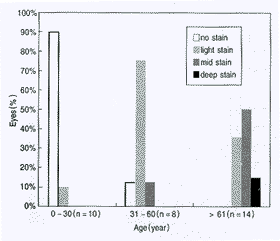
显示染色程度随年龄而增强,条形图代表各种染色程度在不同年龄组中所占的百分比(n=眼数)
图6 不同年龄组中Bruch膜溴-苏丹黑B的染色程度示意图
Fig.6 The histogram demonstrates the different staining intensity of bromine Sudan black B in Bruchs membrane of three age groups.The vertical bars represent the percentage of eyes with a particular staining intensity within each age group.The graph highlights an increasing staining intensity with age(n=number of donor eyes)
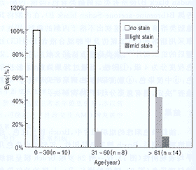
显示染色程度随年龄而增强,条形图代表各种染色程度在不同年龄组中所占的百分比(n=眼数)
图7 不同年龄组中Bruch膜溴-丙酮-苏丹黑B的染色程度示意图
Fig.7 The histogram demonstrates the different staining intensity of bromine acetone Sudan black B in Bruchs membrane of three age groups.The vertical bars represent the percentage of eyes with a particular staining intensity within each age group.The graph highlights an increasing staining intensity with age(n=number of donor eyes)
上一页 [1] [2] [3] 下一页 |
