|
2 结果
2.1 培养RPE的一般特征 与文献报告结果相一致[3],汇合单层的RPE细胞呈多边形。偶联罗丹明的毒伞素标记的肌动蛋白主要位于细胞周边、细胞间连接复合体处(图1a,b)。
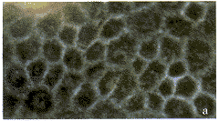 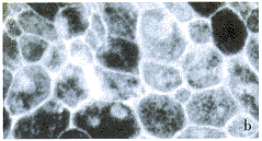
a.生长两周的RPE细胞单层的形态 ×125;b.罗丹明—毒伞素标记技术显示邻近细胞的肌动蛋白丝在细胞与细胞连接处形成单线外观 ×250图1 培养RPE细胞的相差显微像和免疫细胞化学标记后肌动蛋白丝的分布模式
a.Morphology of RPE cell monolayer grown for 2 weeks ×125;b. rhodamine-phalloidin labeling shows actin microfilaments of adjacent cells forming a single line at the site of the cell-to-cell junctions ×250
fig.1 Phase-contrast photomicrograph of cultured RPE cells and the distribution pattern of actin microfilaments by immunocytochemistry.
2.2 基因组DNA电泳 与RPE细胞共培养的抗原特异性活化淋巴细胞,在共培养24小时后,开始出现凋亡征象,48小时可见典型的阶梯状改变,与标准分子量DNA参照物比较为180~200碱基对(base pair,bp)不同倍数的DNA片断(图2)。而与RPE细胞培养上清共培养的抗原特异性活化淋巴细胞、单纯培养的抗原特异性淋巴细胞和与RPE共培养的静止淋巴细胞均未见显著的DNA断裂现象。
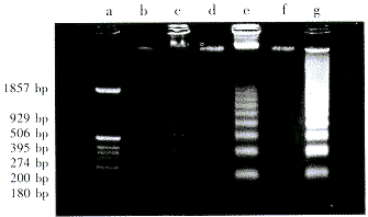
泳道a.分子量标准;b.与RPE培养上清接触72小时的活化淋巴细胞的DNA;c,e,g.分别与RPE共育24,48,72小时的活化淋巴细胞的DNA;d.与完全培养基接触72小时的活化淋巴细胞的DNA;f.与RPE共育72小时的静止期淋巴细胞的DNA图2 与RPE共育的活化淋巴细胞的基因组DNA凝胶电泳 淋巴细胞的核小体间DNA断裂
lane a.Molecular size marker;Lane c,e,g.DNA was isolated from activated lymphocytes cocultured with RPE cells for 24,48,72 hours respectively.Lane b.DNA was isolated from activated lymphocytes exposed to PRE cell cultural supernatant for 72 hours;Lane d.DNA was istolaed from activated lymphocytes exposed to complete media for 72 hours;Lane f.DNA was isolated from resting lymphocytes cocultured with RPE cells for 72 hours
fig.2 Internucleosomal DNA fragmentation of activated lymphocytes cocultured with RPE cells
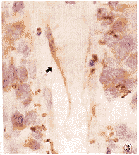
2.3 细胞原位凋亡标记 与RPE细胞共培养24小时的抗原特异性淋巴细胞,其少量的细胞核被TdT酶标记,细胞核呈紫蓝色(图3),随共培养时间延长,被标记的细胞数量有增多趋势。共育24,48,72小时的凋亡细胞的数量分别为(6.7±5.7)%、(9.8±8.8)%和(18.4±18.5)%。而单纯活化淋巴细胞在培养24,48,72小时的标记细胞数量分别为(0.3±0.3)%、(1.3±0.6)%和(3.8±2.5)%;静止淋巴细胞的标记细胞数量分别为(0.2±0.2)%、(0.3±0.5)%和(0.5±0.6)%,与共育组细胞相比均具有统计学显著性差异(P值均小于0.01)。
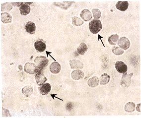
图3 与RPE共育72小时的活化淋巴细胞的显微镜像。细胞核内有蓝紫色颗粒者为凋亡阳性细胞(箭) TUNEL染色 ×250
fig.3 TUNEL stain of activated lymphocytes cocultured with RPE cells for 72 hours(arrows) TUNEL stain ×250
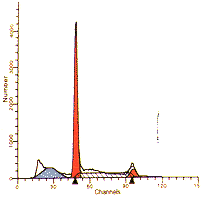
2.4 流式细胞仪检测 与RPE细胞共培养的抗原特异性淋巴细胞:共培养24小时的淋巴细胞,在DNA直方图上,在G1峰左侧出现亚二倍体细胞群的峰形,随着共培养时间的延长,该细胞群的峰形逐渐变宽。凋亡细胞定量结果显示:共培养24小时,凋亡细胞占5.95%;共培养48小时,凋亡细胞占9.38%;共培养72小时,凋亡细胞占17.95%(图4a,b)。其它各组抗原特异性淋巴细胞和静止的淋巴细胞均未见显著的凋亡细胞峰,凋亡细胞定量结果显示:共培养24~72小时,凋亡细胞仅占0.37%~3.86%。
 |
 |
| S-Phase Assessment:
Tissue Type:Lymphocytes
Model type:Diploid
Diploid S:High
Calculated p-value:p<0.01
S-phase Boundaries |
S-Phase Assessment:
Tissue Type:Lymphocytes
Model type:Diploid
Diploid S:High
Calculated p-value:p<0.01
S-phase Boundaries |
 |
a.与RPE细胞共育24小时的淋巴细胞,凋亡细胞占5.95%;b.与RPE细胞共育72小时的淋巴细胞,凋亡细胞占17.9%图4 RPE诱导活化淋巴细胞凋亡的流式细胞仪分析
a.The histogram show lymphocytes cocultured with RPE cells for 24 hours,the apoptosis cells account for 5.95%;b. The histogram show lymphocytes cocultured with RPE cells for 72 hours,the apoptosis cells account for 17.9%
fig.4 Flow cytometry analysis of RPE cell-induced apoptosis of activated lymphocytes
上一页 [1] [2] [3] 下一页 |
