脂质体介导基因转染活体眼组织的研究
眼科研究 2000年第3期第18卷 实验研究
作者:杨桦 张晰 管红兵 王青英 黄璐璐
单位:杨桦 张晰 王青英 黄璐璐(200080 上海市第一人民医院眼科上海市眼科研究所);管红兵(中心实验室)
关键词:眼;转染;脂质体
摘要 目的 了解活体内不同途径应用脂质体转染外源基因在眼组织的分布情况。方法 将包有β-半乳糖苷酶基因质粒的脂质体DOSPER,分别采用玻璃体腔内注射、尾静脉注射、表面点药及球后注射到小鼠体内,2h,1,2天、1,2,4周后用β-gal染色法观察β-半乳糖苷酶基因在眼组织内的分布情况。结果 玻璃体腔内注射、尾静脉注射、表面点药及球后注射4组的小鼠眼球组织包括角膜、虹膜、睫状体、视网膜,均有β-半乳糖苷酶基因表达,除表面点药组外,其他3组小鼠脉络膜也有β-半乳糖苷酶基因表达。用药1天后开始出现β-半乳糖苷酶基因阳性表达,持续达1个月以上。结论 脂质体能有效、稳定地转染外源基因到活体眼组织的角膜、虹膜、睫状体、脉络膜、视网膜。
分类号 R394.3
Experimental observation of gene transfer with liposome to murine ocular tissues by different routes of administration
Yang Hua,Zhang Xi,Guan Hongbing
(Department of Ophthalmology,Shanghai First People’s Hospital,Shanghai Eye Research Institute,Shanghai 200080)
Abstract Objective To compare different routes of administration on the ability of liposomes carrying an exogenous gene to introduce that gene into murine ocular tissues.Methods Plasmid DNA(pGME-7Zf(+)) with beta-galactosidase gene (DOSPER) was applied topically to eyes or injected into vitreous,tail vein,or retro-orbital region of adult mice.Control and treated mice eyes were enucleated at 2h,1d and 2d,1 week,2and 4weeks post-injection or topical application.Gene expression was detected by enzymatic color reaction using beta-gal stain as a substrate in frozen sections of enucleated eyes.Results Cell positive for beta-galactosidase expression showed blue staining of enzymatic color reaction in tissue sections.Liposome-mediated gene transfer was detected in murine cornea,iris,ciliary body and retinal ganglion cells,photoreceptors,and retinal pigment epithelium using all four routes of administration Injection into vitreous,tail vein and retro-orbital region allowed transfer of the gene into the choroid.The gene could not be transferred into murine choroid by topical application.Gene expression in murine ocular tissues was observed after 1d following injection or topical application.The expression lasted at least 1month.Blue staining indicating an enzymatic color reaction was not observed in all of control eyes.Conclusion Efficient and stable transfer of functional genes could be achieved by liposome delivery into the cornea,iris,ciliary body,choroid and retina of mice.
Key words ocular transfer liposome
目前已发现许多致盲性遗传眼病存在不同基因突变,尤其是已查明的视网膜变性疾病的基因突变多种多样[1],基因转移技术是体细胞水平更正基因突变的有效方法[2]。研究表明以病毒作为载体,可有效地转移正常的功能基因到活体眼组织,通常使用的病毒有逆转录病毒、腺病毒及单纯疱疹病毒等[3,4]。但是在眼部应用上述病毒,有可能导致其他的病毒疾病,也就是说安全性不可靠。脂质体是一类非病毒制剂,作为基因转移的载体具有低毒性、相对价廉和使用方便等优点,而且具有一定的转染效率。本研究以脂质体作为载体,通过玻璃体腔内注射、尾静脉注射、表面点药及球后注射等多种途径给药,观察不同时期小鼠眼组织外源基因转染情况,为应用脂质体进行体细胞基因治疗奠定基础。
1 材料和方法
1.1 材料 脂质体1,3-Di-Oleoyloxy-2-(6-Carboxy-spermyl)-propylamid(DOSPER)和β-gal染色试剂盒为德国BOEHRINGER MANNHEIM公司产品;携带β-半乳糖苷酶报导基因的重组质粒pGME-7Zf(+)为华美公司产品。体重40g左右的昆明小鼠。美国Becton Dickinson公司的30G1/2号针头,德国Leica显微镜。
1.2 方法 戊巴比妥钠腹腔内注射麻醉小鼠,小鼠分为5组:1组:玻璃体腔内注射DOSPER与pGME-7Zf(+)质粒的混合液;2组:尾静脉注射DOSPER与pGME-7Zf(+)质粒的混合液;3组:眼球表面滴用DOSPER与pGME-7Zf(+)质粒的混合液;4组:眼球后注射DOSPER和pGME-7Zf(+)质粒的混合液;5组为对照组,上述4种途径分别使用DOSPER或pGME-7Zf(+)或生理盐水或空白对照。


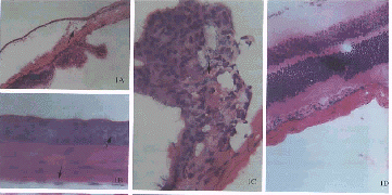
图1 对照组的角膜(A)、虹膜(A,F)、睫状体(A)和视网膜(A,D)。玻璃体腔内注射DOSPER与pGME-7Zf(+)质粒混合液后1周基因转移情况:角膜上皮细胞和内皮细胞(B)、睫状体(C)、虹膜(G)、视网膜节细胞层、光感受器层及色素上皮细胞(E)均可见β-半乳糖苷酶基因的阳性表达 图2 尾静脉注射DOSPER与pGME-7Zf(+)质粒混合液后1周视网膜基因转移情况:脉络膜(a)、视网膜色素上皮细胞(b)、光感受器(c)及节细胞(d)均可见β-半乳糖苷酶基因的阳性表达 Fig.1
Cornea(A),iris(A,F),ciliary body(A) and retina(A,D) in the normal control group.Gene transfer 1week after the injection of DOSPER and pGME-7Zf(+) into the vitreous.Beta-galactosidase expression is
seen in the corneal epithelial cells and endothelial cells(B),ciliary body(C),iris(G),retinal ganglion cells,photoreceptors and pigment epithelial cells(E) Fig.2 Gene transfer 1week after the injection of DOSPER and pGME-7Zf(+) into the tail’s vein.Beta-galactosidase expression is seen in the choroid(a),retinal pigment epithelial cells(b),photoreceptors(c) and retinal ganglion cells(d)
用药后2h,1,2天、1,2,4周分别摘取上述5组小鼠眼球,立即冰冻切片,固定,PBS冲洗,β-gal染色,PBS冲洗,HE染色,甘油或中性树胶封片。显微镜下观察并照相。
[1] [2] 下一页 |
